Fertility and reproduction (F&R) is declining due to a number of environmental and life style factors. To overcome reduced fertility in vitro fertilization (IVF) or intracellular sperm injection in the oocyte (ICSI) were introduced. In both techniques sperm and oocytes are put together to establish fertilization and embryonal development. These approaches are, however, inefficient with species-specific limitations. The main difference with in vivo fertilization is that during IVF or ICSI the oviduct is bypassed. In vivo the oviduct has a role in binding and activating of incoming sperm cells, in guiding the ovulated oocyte(s) during fertilization, and in early embryonic development.
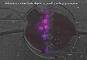
We developed A 3D microfluid oviduct-on-a-chip model in which fertilization and development of embryos can take place in an environment that resembles the in vivo situation. In a microfluidic two perfusion chamber, oviduct epithelial cells form a cell monolayer that are fed at the basolateral side by medium. At the apical side, these cells show secretory and ciliary activity that will allow sperm binding and activation as well as oocytes to become fertilized. The embryos are more alike in vivo embryos when compared IVF or ICSI embryos.
The oviduct-on-a-chip is also used for female reproduction toxicity tests by administering toxic components to the basolateral perfusion compartment and determining indirect effects on sperm cells, oocytes and embryo development at the apical compartment.
All the female materials for this research are obtained from slaughterhouse animals, complying with 3R policy of Veterinary Medicine and Utrecht University to reduce replace and refine animal experiments. F&R is a collaborative project with Farm Animal Health, Equine Sciences, and Companion Animals.